Highlights: Light Scattering Reveals Novel Applications of DNA
Dr. Izhar Medalsy, Product Marketing Manager, Wyatt Technology Corp. - March 29, 2016
Deoxyribonucleic acid (DNA) is an amazingly versatile molecule thanks to its unique structure and stability. Encoding and passing on to subsequent generations all inheritable genetic information, the DNA molecule plays a well-known, pivotal role in the biological world. However, DNA has also been the subject of cutting-edge research in fields outside the usual scope of genetics and biology, from nanotechnology and nanoelectronics to drug delivery and even optogenetics, taking advantage of its highly customizable properties. Light scattering, of course, plays an important part in many of these studies. Read on to find out how!
DNA is not just a genetic biomolecule – it can be a nanotechnological tool as well. Mustafa et al. showcase this capability in their investigation of fluorescent emission from surface-functionalized nanoparticles, controlled by single-stranded DNA (ssDNA) molecules (Mustafa B. et al. (2014). "DNA-length-dependent quenching of fluorescently labeled iron oxide nanoparticles with gold, graphene oxide and MoS2 nanostructures." ACS Appl. Mater. Interfaces 6:12100-10. doi: 10.1021/am503553h). The scientists used Wyatt’s DynaPro® NanoStar dynamic light scattering instrument to monitor the increase in the nanoparticle’s hydrodynamic radius Rh with increasing length of ssDNA, in order to verify attachment and correlation to ssDNA length. In turn, the degree of fluorescence quenching was tuned according to the ssDNA length, providing a glimpse into how hybrid bionanomaterials may be engineered with tunable optical properties.
Antisense oligonucleotides (AONs) hold promise for therapeutic correction of many genetic diseases. However, systemic use of AONs is limited because of poor tissue uptake. Goyenvalle et al. present a new class of AONs made of tricyclo-DNA (tcDNA), designed as a conformationally-constrained oligonucleotide analogue (Goyenvalle A. et al. (2015) "Functional correction in mouse models of muscular dystrophy using exon-skipping tricyclo-DNA oligomers." Nat. Med. 21: 270-5. doi: 10.1038/nm.3765). This DNA variant displays unique pharmacological properties and unprecedented uptake by many tissues including the heart and the brain, making tcDNA-AON chemistry particularly attractive as a potential future therapy for Duchenne muscular dystrophy and other neuromuscular disorders.
SEC-MALS studies utilizing Wyatt’s DAWN® light-scattering instrument and Optilab® helped elucidate the mechanism behind the enhancement pharmacologic performance, indicating that tcDNA spontaneously forms well-defined nanoparticles ranging in size from 40 to 100 nm (rms radius, Rg). This self-associative property, supported by MALS as well as nanotracking analysis (NTA) and electron microscopy, imitates features of transfection reagents and nanoparticle delivery systems that typically exhibit good cellular uptake.
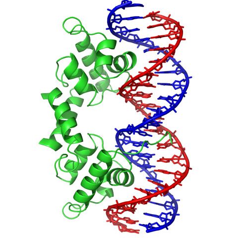
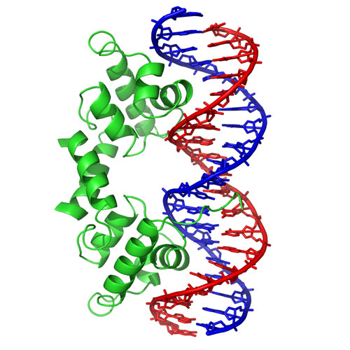
Another exciting development utilizing DNA is optogenetics, a biological technique which involves the use of light to control cells in neurons and other living tissues. The design of synthetic optogenetic tools that allow precise spatiotemporal control of biological processes has developed rapidly over the last few years. In a recent publication, Heintz and Schlichting describe the mechanism of light-dependent DNA binding of the light-oxygen-voltage (LOV) transcription factor from the Phaeodactylum tricornutum diatom (algae) (Heintz and Schlichting (2016). "Blue light-induced LOV domain dimerization enhances the affinity of Aureochrome 1a for its target DNA sequence." eLife 5). SEC-MALS using a DAWN, and other DNA-binding experiments, revealed a light-sensitive interaction between the LOV and DNA-binding domain, wherein exposure to blue light shifts the monomer-dimer equilibrium towards increasing molecular weight. With a recovery time constant on the order of hundreds of seconds, the effect is sufficiently long-lived to be observed in SEC separations, while the equilibrium itself is rapid and exhibits a shift in molar mass, correlated to concentration, across the eluting peak. Aureos’ unique combination of different functionalities offers new design strategies for molecular switches, to control biological processes with unprecedented spatial and temporal precision.
Increasing insight into the structure and functionality of DNA has led to the development of structural DNA nanotechnology, wherein DNA not only encodes biological information but also serves as a polymeric, structural material. By means of its molecular recognition properties, DNA can be manipulated to self-assemble into higher order structures with unique nanobiotechnology applications ranging from sensing to nanocarriers and drug delivery vehicles. Tang et al. utilized a DynaPro dynamic light scattering detector alongside atomic force microscopy (AFM) to characterize the size distribution of the hydrodynamic radii and micellar core heights, respectively (Tang L. et al. (2014), “Enzymatic Polymerization of High Molecular Weight DNA Amphiphiles That Self-Assemble into Star-Like Micelles.”, Adv. Mater. 26:3050-54. doi: 10.1002/adma.201306049). DLS showed great agreement with dissipative particle dynamics simulations, stressing once again the strength of light scattering in measuring particle size.
Whereas MALS and DLS are used regularly to characterize DNA and its interactions with common binding partners such as DNA-binding proteins, it is gratifying for us folks at Wyatt Technology to know that our light scattering instruments are also helping to push the boundaries of DNA-based nanotechnology and nanomedicine. If you have published an article about characterization of DNA using Wyatt instruments (or any other macromolecule or nanoparticle, for that matter), please let us know so we can share the information in our online bibliography at www.wyatt.com/Bibliography. Send your preprint to: publication@wyatt.com